Classification of Diabetic Retinopathy : Background Retinopathy
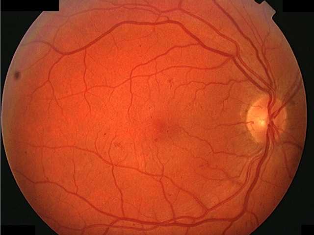
Background retinopathy (BGR) or non-proliferative retinopathy always predates proliferative retinopathy.
Initially BGR consists of microaneurysms only...
progressing to microaneurysms with small haemorrhages (dot and blot haemorrhages)...
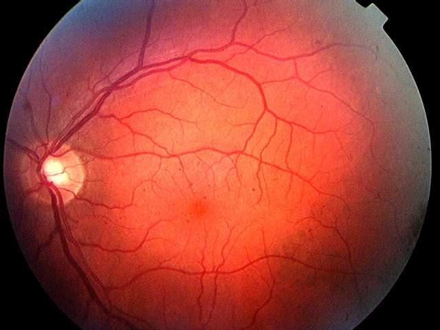
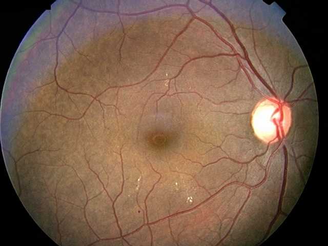
and flame-shaped haemorrhages (labelled 1 in the image below) which are also associated with hypertension...
The image above also shows a crescent on the temporal aspect of the optic disc (labelled 2) which is a normal variant.
Maculopathy
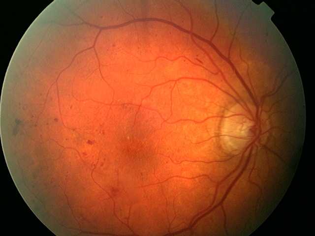
Hard exudates are also consistent with the designation of background retinopathy...
However, where these changes - both haemorrhages...
and hard exudates...
are within the macular area, the picture is designated as ‘Diabetic Maculopathy’ so as to highlight the potential sight-threatening nature of this condition.
Diabetic maculopathy is classified into focal, diffuse and ischaemic types although for physicians this is a rather academic exercise since all patients will require a formal ophthalmic review. Visual loss is due to macular oedema (which can only be viewed by binocular stereoscopic slit lamp examination) and/or ischaemia. Differentiation of these components is usually achieved by fluorescein angiography.
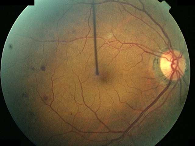
Cotton Wool Spot
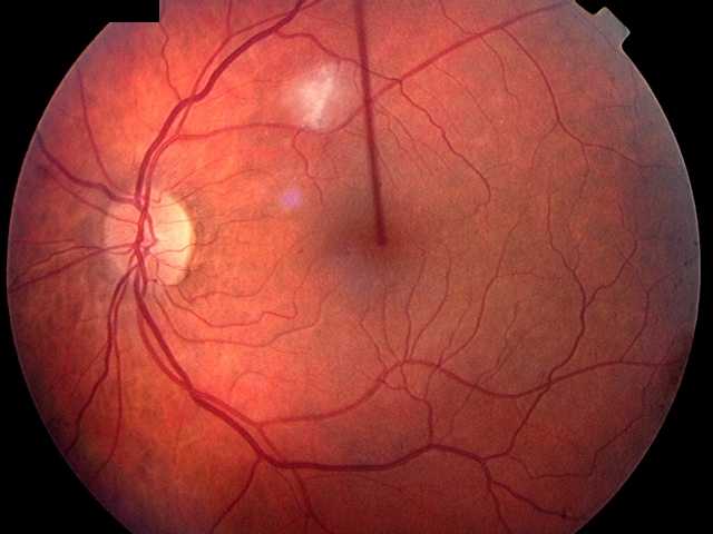
An occasional cotton wool spot and/or mild venous dilatation (which is so subjective that it cannot usually be seen) is consistent with background retinopathy.
Steve Bain, Jon Gibson, Paul Dodson, Graham Sedgwick