Normal Features : General
This image shows a normal retina of a left eye. The retina is made up of two layers, the outer retinal pigment epithelium and the inner neuro-sensory layer.
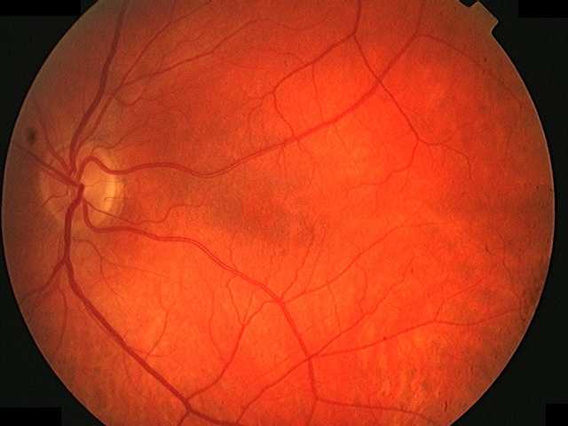
The optic disc (1) is entry of optic nerve fibres, it measures approximately 1.5mm in diameter.
The macula (2) is the central area of the retina. Within this lies the fovea (3), a central depression in the macula which is approximately 1.5mm in diameter. Using direct ophthalmoscopy, the fovea is seen as a light reflection (termed ‘reflex’). On these images, the precise location of the fovea can only be estimated. The macula is said to extend approximately 2 disc diameters from the fovea.
The macula is the specialised area of maximal spatial, temporal and colour discrimination due to the high concentration of cone photoreceptor.
Steve Bain, Jon Gibson, Paul Dodson, Graham Sedgwick